Neuroimaging to the Rescue: One woman's cancer journey reveals a decades-long quest

It began with the sound of a heartbeat. Ingrid Youmans—a vibrant, middle-aged consultant, designer, and mother of two—swore she could hear the quiet rush of blood behind one ear, pulsing with the rhythm of her heart. It was subtle at first. Infrequent. So, she dismissed it, busy mom that she was, chalking it up to just one more of those weird things bodies do in their 40s. But the sound persisted, growing more frequent as the weeks churned. She decided to mention it at her annual checkup.
It happens, Youmans’ doctor told her, like when you bend over and stand up straight again. But Youmans insisted this experience was different. It wasn’t a change in blood pressure responding to some action taken. She could be lying in bed, perfectly still, and begin to hear the beating of her heart, like an urgent whisper in her ear. The doctor ordered an MRI, just in case.
“She basically saved my life with that one decision,” Youmans says of her physician.
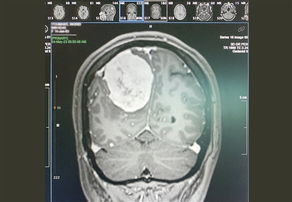
When Youmans went in for her scheduled MRI, she quickly received some disquieting news. “As soon as my head cleared the machine and they took the headphones off,” she recalls, “the MRI tech looked at me and said, ‘We’re admitting you to the ER.’” Inside Youmans’ skull was a golf-ball sized grade I meningioma—a tumor of the membranous linings of the brain—and it was growing.
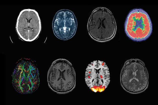
According to Guoying Liu, director of the Division of Applied Science and Technology at the National Institute of Biomedical Imaging and Bioengineering (NIBIB), neuroimaging is a powerful noninvasive tool used to manage patients with possible brain cancer. Doctors tend to favor three main imaging modalities to see what’s going on inside the brain: computed tomography (CT), positron emission tomography (PET), and magnetic resonance imaging (MRI).
Computed tomography is often the first modality used to check a patient presenting with a brain tumor, Liu says, because CT is fast, cost efficient, and provides better spatial resolution than MRI. But MRI is the primary imaging modality routinely used in the care of brain tumor patients due to its ability to provide structural, functional, and metabolic information noninvasively. “PET scans can be very specific for detection of tumors, and they are highly sensitive, showing metabolic changes that occur at a cellular level,” she says. But the downside to PET and CT imaging is that they expose the patient to radiation. MRI scans, on the other hand, do not have any radiation impact.
MRIs have been used by doctors since the 1970s to take snapshots of the body’s internal structures. Youmans, who finds humor even in the darkest of times, laughs when she thinks about her experience with the machines. “An MRI feels like an actual practical joke being played on you,” she says. “You’re in a tube where there’s just banging and, like, someone is sawing, someone is playing a super uncomfortable xylophone–it doesn’t make any sense. What’s happening around you that could possibly achieve a detailed picture of anything?”

The large, humming scanners of an MRI machine use a series of superconducting coils submerged in a frigid liquid helium bath to measure quantum information from the body. At rest, the machine emits a strong magnetic field, overpowering the natural mishmash of polar directions of the body’s hydrogen protons, aligning them with the machine’s magnetic field. Then, a pulse of radio waves is sent from the coils at a specific frequency, jostling the spinning protons 90 or 180 degrees from the machine’s field alignment. As the spins of these protons relax, the scanner measures the time it takes their polarities to realign with the larger magnetic field, interpreting their signals into a grayscale graphic of tissues. Tumors like meningiomas may then display on a screen as a bright white mass in a field of black and gray.
According to Chetan Bettegowda, professor of neurosurgery and director of the Meningioma Center at the Johns Hopkins University School of Medicine, meningiomas are the most common type of primary brain tumor. “Some autopsy studies have shown that even up to one percent of the population may have a meningioma and never know about it,” he says. And while most meningiomas are benign like Youmans’, they can still wreak havoc on the brain.
“When most people think about benign tumors, they assume it’s something that’s not going to cause harm,” says Bettegowda. “We know that really, any sort of abnormal process—whether it’s benign or malignant—that’s growing, unfortunately, can have very significant consequences on neurological function. And if it gets extreme enough, on survival, as well.”
Although tumors attached to the meninges have been reported since the mid-1800s, it wasn’t until neurology giants Louise Eisenhardt and Harvey Cushing literally wrote the book on them in 1938 that the term “meningioma” became medicinal canon. At that time, meningiomas seemed rare—they weren’t usually discovered until a person’s ability to function was greatly impaired. Doctors typically operated using just their knowledge of brain function, the neurological symptoms presented by their patients, and any observable defects found. Death during and post-surgery was frequent.
“The very first [patients with] tumors taken out by Harvey Cushing and others had very high mortality rates, secondary to things like infection and neurological injury and anesthetic complications and bleeding risks,” says Bettegowda. “A hundred-plus years ago, the surgeries were dire—extreme opportunities to try to preserve life knowing that the consequences might hasten the demise.” Thankfully, doctors today are able to peer inside the body to capture as much information about these growths before ever touching a knife.
After her MRI, Youmans was escorted to a bed in the ER where she awaited full results of the scan. When the attending physician came in, she explained what the MRI found: a large, metabolically active mass attached to the outer layer of her meninges. The tumor had disrupted her blood-brain barrier, causing fluid to accumulate and build pressure in her skull. This increased pressure was irritating the optic nerve, causing the nerve sheath to thicken. The pressure was also injuring the surrounding healthy tissue, compressing it and pushing it aside. The tumor had pushed so much aside, in fact, it had shifted the midline of her brain by almost a centimeter.
Later that day, Youmans was whisked off to a larger hospital where neurosurgeons could remove the tumor. But before that could happen, she needed to be scanned with X-rays by a CT machine.
X-rays, discovered in 1895 by Willhelm Conrad Röntgen, have been used in medical imaging since the year after their discovery. The ghostly images can reveal, for example, breaks, spurs, and other aberrations in bones. Soft tissues, on the other hand, all have similar densities so that on X-ray images, they mostly appear as undifferentiated gray blobs. For this reason, X-rays weren’t widely used by neurosurgeons for decades. Then, in the mid-1910s, Cushing protegee and Johns Hopkins neurosurgeon Walter E. Dandy realized that air appears as bright spots on developed X-ray images, so he applied his discovery to his practice.

A photo showing the MRI results of Youmans' meningioma. The total size of the tumor, after surgery, was approximately 7 cm x 5 cm x 3 cm. Photo credit: Ingrid Youmans, 2023
A few years later, Dandy invented procedures like ventriculography and pneumoencephalography (PEG) that replaced cerebrospinal fluid with air, thereby increasing contrast between soft tissues on X-ray images. Doctors could now see some lesions or blockages in the brain, which could then guide surgical plans. Introducing air into spinal fluid, however, caused extreme pain and could lead to serious side effects like meningitis or even death. More accurate and less invasive tests like CT finally replaced those procedures in the early 1970s.
A CT scanner is a large, donut-shaped machine with a table for the patient to lie on that slowly passes through the donut’s opening. An X-ray emitter and detector on opposite sides of the donut spin rapidly around the patient, building a one-slice-at-a-time view of the person’s insides as their body moves through the opening.
To visualize blood vessels in CT scans, an angiogram is often required. An iodine-based contrast dye is injected into the bloodstream to block X-rays from the scan. A computer then takes this information and creates a map of the blood vessels in the brain, allowing doctors to pinpoint arteries that might be feeding tumors. Next, with a catheter, they travel through the artery and stop the blood flow with an embolization, (from Greek embolos: wedge-shaped stopper) thereby starving the tumor cells of nourishment.
“It’s the most-odd sensation ever,” Youmans says of the embolization. “I could feel when [the technician] clipped. It felt like he inserted two chip clips into my brain—there was this snapping noise, and I could hear it inside my brain!”
Once Youmans’ meningioma was cut off from most of its blood supply, and further imaging verified there were no additional tumors, her surgery was scheduled for a few days later. She used those days to draw or spend time with family, trying to process what was happening.
“It was really nice to have people there, but every time someone said goodbye, it was like ‘How do you say goodbye?’ Everybody knows this could be it,” Youmans says. It was especially hard with her eight-year-old son, Jude. “That was the first time I think he’s seen me as not invincible.”
Youmans wrote letters to her family, just in case, and her kids drew her pictures to keep her spirits up. On the day of her surgery, the craniotomy began around 8 am. With the help of a surgical navigation system called StealthStation that uses infrared light to tie a sensor wand with an MRI map of Youmans’ brain, neurosurgeon Angelique M. Ruff slowly, delicately, deliberately excised the meningioma. By 5:30 pm, Youmans was being prepped for release from the operating room, tumor-free.
While Youmans’ meningioma was removed relatively quickly, not all tumors can be handled that easily. Some grow deep inside the brain, where applying concentrated beams of gamma radiation may be the only solution to destroying them. But even radiosurgery requires a good understanding of what’s going on inside to ensure accuracy and successful treatment. Thankfully, medical imaging is getting better and better.
NIBIB’s Liu says that much of the excitement in intracranial imaging is happening with technology advances in the MRI realm. For instance, perfusion MRI, which can be performed with or without contrast agents, allows imaging of blood circulation. Diffusion-weighted and diffusion-tensor MRI measure composition and structure of tissue. And MR spectroscopy looks at a tissue’s chemical makeup.
“NIBIB, in the past 10 years or so, has been supporting quite a number of institutions in their development of advanced methodologies,” says Liu, who gives the example of chemical exchange saturation transfer (CEST) MRI and hyperpolarized carbon-13 MR spectroscopic imaging.
CEST MRI, like traditional MRI, detects the relaxation of proton spins in water molecules. It differs, though, by continuously exciting other chemical protons through different frequencies, allowing protons to transfer to water molecules until a magnetic saturation point is reached, and then measuring the proton spin relaxation. Hyperpolarized carbon-13 MR spectroscopy, however, is more like a chemical “tracer,” using thermal modifications of enriched C-13 isotopes to reveal information about things like metabolism, chemical reactions, or the functioning of specific organs.
“These new methodologies really open the door for many kinds of cancer imaging, including brain cancer,” says Liu.
Several months after Youmans’ procedure, she described feeling like herself again, though she tires easily from a full day of work, sometimes forgetting things or writing notes twice. “I think it’s clear my brain is still relearning some things,” she says.
Youmans says she can still occasionally hear her heartbeat by her ear. Her doctors reassured her that it’s nothing alarming—brain rewiring takes time, but not everything returns to normal. “So, I hear it once in a while and it’s a healthy reminder,” she says. A healthy reminder of her ordeal. Of her family. Of the people and technology that saved her life.
Rachel Lense is a freelance science writer, poet, and artist living in the greater Washington, DC area. She loves to tell stories at the intersection of science, technology, and culture, and emphasizes the dual roles history and language tend to play therein.


