Static real-time capture of 3D microscopy images
Two-dimensional widefield microscopy provides basic dynamic information about live biological specimens. However, it provides only a partial representation of the 3D biological processes and may be incomplete or even misleading. Current techniques, such as widefield, confocal, structured-illumination, or light-sheet microscopy cannot capture the 3D structure of a specimen in a single frame. In contrast, in 2D widefield microscopy, a stack of 2D depth images of the sample are recorded and a 3D digital image is computed from them. The different depth images are typically recorded with axial mechanical scanning. But mechanical movement could damage the sample, cause it to vibrate and hence introduce image distortions, or slow down image acquisition, which would make it impossible to record highly dynamic biological processes. The trivial solution is to use digital holographic microscopy, which permits the 3D complex distribution scattered by the sample to be rendered from a single frame.1 However, this system operates coherently and makes fluorescence imaging impossible.
 We have investigated using an electrically addressable liquid lens (LL) to acquire images at different depths. The lens is based on electrowetting technology: how a drop of water spreads on an electrically insulating surface can be modified by accumulating charge at the base of the drop. The optical power of the resulting LL can be tuned by an applied voltage.2 In 2010, our group proposed using an LL for parallel dynamic focusing of images obtained through an array of microlenses.3 More recently, other groups have applied LL technology in microscopy.4, 5
Our proposal is to insert an LL at the aperture stop of a widefield microscope, which is arranged as the telecentric coupling between a high-numerical-aperture (NA) infinity-corrected microscope objective and a low-NA tube lens.6 The insertion of the LL enables the axial position of the object plane to be controlled by the voltage while preserving the telecentric nature of the microscope, its lateral magnification, and the position of the image plane. In our proof-of-concept experiment we inserted an afocal relay into the optical path (see Figure 1). It is easy to show that in such a case the relation between the optical power of the liquid lens, PLL, and the induced displacement, Δ, of the object plane is Δ=fob′2 MRel2PLLwhere MRel=−f ′ R2/f ′ R1 is the lateral magnification of the relay and fob′ is the focal length of the microscope objective.

To verify the accuracy of the system, we first measured the lateral point spread function (PSF) for different values of PLL. To perform the experiment, we inserted an LL ARCTIC 39N0 (VARIOPTIC) in our optical microscope composed by a 50×/0.55 microscope objective, a relay with focal lengths f ′ R1=300mm and f′R2=100mm, and a tube lens of f′TL=200mm. In Figure 2 we show the measured PSFs, which demonstrate that the LL hardly affects the PSF shape and size.

Once we had verified that the LL produces no significant worsening of lateral PSF, we performed an imaging experiment in which we obtained a stack of depth section images of a cleaning-lens tissue paper. These sheets have a low thread density, and are about 100μm thick. Figure 3 shows a set of depth images obtained with the proposed setup and without mechanical movement.
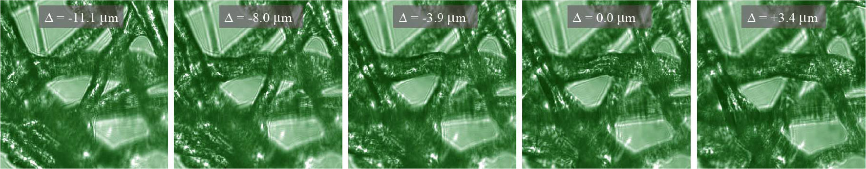
In summary, we have shown that tuning the LL applied voltage enables the real-time capture of a stack of depth images of 3D specimens with constant magnification and resolution. The main advantage of this technique is performing fast axial scanning free of mechanical vibrations. We are now working on a prototype in which the liquid lens is inserted, directly, at the objective aperture stop. We are also preparing user-friendly, fast software for processing and visualizing the 3D images.
This work was supported by the Spanish Ministry of the Economy and Competitiveness (DPI2012-32994) and also by the autonomous government of Valencia, Spain (PROMETEOII/2014/072).
University of Valencia
Manuel Martínez-Corral is a full professor of optics and co-leader of the 3D Imaging and Display Laboratory. He received a PhD in physics from the University of Valencia in 1993. In 2010, he became an SPIE Fellow.
Ana Doblas received a BSc and MSc in physics from the University of Valencia in 2010 and 2011, respectively. Since 2009 she has been working at the 3D Imaging and Display Laboratory where she is currently a PhD student and holds a predoctoral fellowship. Her research interests include 3D optical microscopy and image formation theory.
Emilio Sánchez-Ortiga received an MSc and PhD in physics from the University of Valencia in 2009 and 2014, respectively. He has published 11 manuscripts in major journals and authored over 30 communications in scientific conferences. He currently holds a postdoctoral contract, and his main research interests are 3D microscopy, including confocal scanning microscopy, structured illumination microscopy, and digital holographic microscopy.
Jorge Sola-Pikabea received a BSc in physics from the University of Valencia, Spain, in 2014 and has been a master's student at the 3D Imaging and Display Laboratory since completing his degree. His research interests include 3D microscopy.
Genaro Saavedra is a full professor of optics and co-leader of the 3D Imaging and Display Laboratory at the University of Valencia. In 1996 his doctoral research was honored with an Extraordinary Award. His research interests include microscopic and macroscopic 3D imaging. He has published more than 70 papers and given more than 20 invited talks on these topics.