Evaluating Kidney Function
Article in Journal of Biomedical Optics on optical assessment method.
A new optical technique developed by American researchers promises to improve accuracy and reduce costs of real-time assessment of kidney function.
An open-access article published in May in the Journal of Biomedical Optics explores the use of multimodal autofluorescence and light scattering to evaluate functional changes in the kidneys after ischemic injury. Conditions such as accumulated arterial plaque or blood clots restrict the flow of oxygen and glucose to organs, and prolonged periods of such ischemia can compromise function.
In “Predictive assessment of kidney functional recovery following ischemic injury using optical spectroscopy,” the authors report on their evaluation of various optical signatures to predict kidney viability and suggest a noncontact approach to provide clinically useful information in real time.
While other work in this area uses expensive multiphoton and laser-based techniques, the authors lowered expenses by switching to camera-based imaging.
No real-time tool is in current use to measure the degree of ischemic injury incurred in tissue or to predict the return of its function. Doctors unable to decisively determine tissue functional status run the risk of increasing patient morbidity and mortality if they transplant dysfunctional tissue. In addition, they may discard much-needed functional kidney tissue because they are unable to assess its viability.

The study was authored by Rajesh Raman, a scientist at Lawrence Livermore National Lab, and coauthors Christopher Pivetti, a research lab supervisor at the University of California, Davis Medical Center; Rajendra Ramsamooj, a transplant expert from California Northstate University College of Medicine; Christoph Troppmann, a transplant surgeon at UC Davis; and Stavros Demos, a senior scientist at the Laboratory for Laser Energetics at University of Rochester.
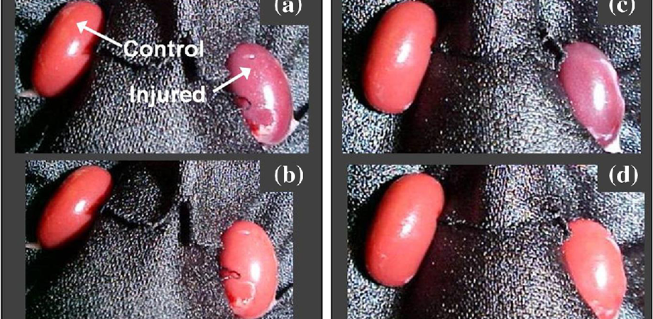
The authors acquired autofluorescence images of rat kidneys in vivo under 355, 325, and 266 nm illumination. They collected light-scattering images at the excitation wavelengths while using a relatively narrow band light centered at 500 nm.
The images were simultaneously recorded using a multimodal optical imaging system. Researchers then analyzed the recorded signals to obtain time constants, which were correlated to kidney dysfunction as determined by a subsequent survival study and histopathological analysis.
Analysis of the light-scattering and autofluorescence images suggests that variations in tissue microstructure, fluorophore emission, and blood absorption spectral characteristics, combined with vascular response, contribute to the behavior of the recorded signals. These are used to obtain tissue function information and enable the ability to predict post-transplant kidney function.
This information can also be applied to the prediction of kidney failure when visual observation cannot, almost immediately following an injury.
Reviewers of the article suggested other promising applications for future development and envisioned the approach being used as a screening tool for assessing kidney viability prior to transplant. In particular, they said, these cost-effective screening methods could benefit healthcare in developing countries.
Multimodal imaging also has provided insights into other physiological events that may occur during ischemia and reperfusion.
“This work's exceptional value lies in the realization of a workable practical system that has excellent potential to be adopted in field situations,” said SPIE Fellow Andreas Mandelis of University of Toronto, a journal associate editor.
Source: dx.doi.org/10.1117/1.JBO.22.5.056001
- Have a question or comment about this article? Write to us at spieprofessional@spie.org.
- To receive a print copy of SPIE Professional, the SPIE member magazine, become an SPIE member.


