Handheld, lensless microscope identifies malaria parasites
Optical microscopy is an indispensable tool in science and medicine. However, the size and cost of conventional optical microscopes limit their use in rugged field-settings or in remote areas to assist in tasks such as medical tests, disease diagnostics, or monitoring of water quality.
In recent years, there has been widespread interest in reducing the size and cost of microscopes and making them more suited for point-of-care and telemedicine applications, or for tackling global health problems. These approaches range from miniaturizing traditional microscope designs and attaching them to cell phones, to eliminate lenses altogether and using digital sensors in very close proximity to the specimen.1–6
We followed the latter approach to create a field-portable lensless optical microscope that can image large sample areas with sub-micron resolution. This compact microscope is based on an imaging technique termed ‘partially-coherent digital in-line holography.’ This method uses simple light sources, such as light-emitting diodes (LEDs) and off-the-shelf digital image sensors without the need for any lenses or other sensitive or bulky optical components. We also use a digital image processing technique called pixel super-resolution to convert multiple low-resolution microscope images to a single high-resolution one.

Figure 1 shows a photograph and a schematic diagram of our portable holographic microscope. The device weighs 95g, can be powered by battery or USB, and has a field-of-view (FOV) of 24mm2. The core configuration is made up of a CMOS image sensor. The sample to be imaged is placed on top of the sensor and is illuminated by a multimode optical fiber with a LED butt-coupled to one of its ends.
In this configuration, the light reaching the sensor is either scattered from the object or transmitted unaltered through the sample. The sensor records the intensity of the interference pattern between the scattered and undisturbed light, forming a hologram (see Figure 1). The phase of the light field is lost because the sensor responds only to intensity, but iterative phase retrieval algorithms enable recovery of the phase.6 In turn, this allows the field to propagate back to the sample plane, which enables recovery of a microscopic image of the object in both the amplitude and phase channels.

The disadvantage of this arrangement is that the pixel size of the sensor limits the image resolution.7 To remedy this, we use several multimode optical fibers, each butt-coupled to an individual LED. By sequentially turning on the LEDs, we capture multiple shifted holograms of the stationary object located on the chip. This hologram effectively has a smaller pixel size, leading to a higher resolution microscopic image (see Figure 1).8
The proximity of the sample to the sensor in this configuration allows the use of the entire FOV of the sensor as the microscope's FOV. This area can be orders of magnitude larger than that of a traditional microscope. With a useful magnification (×10–×40), our approach is appropriate for imaging applications that require testing a large sample volume, such as looking for rare parasites in water or blood.
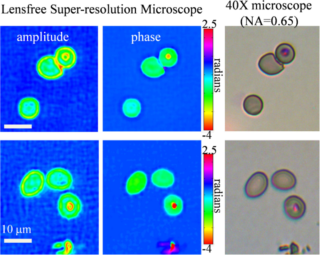
The possible use of this microscope in addressing global health problems is demonstrated in Figure 2, which shows red blood cells infected with malaria parasites. The combination of amplitude and phase images allows the identification of malaria parasites within the red blood cells, making it possible to distinguish healthy blood cells from infected ones. The large FOV of the microscope includes thousands of red blood cells, which can potentially allow automated malaria diagnosis using a single image from this portable microscope. Note that a traditional microscope, due to its limited FOV, would require multiple scans to get a statistically significant number of cells.
In conclusion, lightweight, compact, and cost-effective optical imaging and micro-analysis devices would allow greater accessibility to health care throughout the world. We have described our approach to creating such a device, which is based on lens-free digital holography along with image processing algorithms. We have also shown that our microscope is capable of imaging malaria parasites in human blood. By further improving their performance, reliability, and cost-effectiveness, we aim to automate malaria diagnosis using our field-portable super-resolution microscopes.
University of California
University of California


